中国全科医学 ›› 2022, Vol. 25 ›› Issue (26): 3281-3289.DOI: 10.12114/j.issn.1007-9572.2022.0217

• 论著 • 上一篇    下一篇

透明质酸寡糖通过影响核因子E2相关因子2信号通路促进糖尿病皮肤溃疡创面愈合的机制研究

胡钟元1,2, 张秀华2, 李帅广2,3, 邵华荣2, 刘飞1,2,3,*(), 郭斌1,*()   

  1. 1.121001 辽宁省锦州市,锦州医科大学药学院
    2.250101 山东省济南市,山东省药学科学院 山东省生物药物重点实验室
    3.250012 山东省济南市,山东大学齐鲁医学院药学院
  • 收稿日期:2022-02-25 修回日期:2022-05-10 出版日期:2022-09-15 发布日期:2022-05-19
  • 通讯作者: 刘飞, 郭斌

  • 作者贡献:胡钟元负责实验设计的实施、实验指标的检测以及撰写论文,并进行数据处理、统计学分析、绘制图表;张秀华、李帅广负责进行动物造模以及论文的修订;邵华荣负责最终版本的修订以及论文内容的审校;刘飞、郭斌负责确定文章选题,指导实验设计及论文撰写。 胡钟元,张秀华,李帅广,等.透明质酸寡糖通过影响核因子E2相关因子2信号通路促进糖尿病皮肤溃疡创面愈合的机制研究[J].中国全科医学,2022,25(26):3281-3289.[www.chinagp.net]
  • 基金资助:
    国家重点研发计划(2021YFC2103100)——医用多糖生物制造关键技术及产业化示范; 山东省重点研发计划(重大科技创新工程)项目(2021ZDSYS07)——医药绿色智能制造与新药评价关键技术研究; 济南市"高校20条"资助项目"天然产物新功能发现及生物合成创新团队"(2019GXRC038)

Effect of Hyaluronic Acid Oligosaccharides on Diabetic Cutaneous Ulcer Wound Healing by Activating Nrf2 Signaling Pathway

Zhongyuan HU1,2, Xiuhua ZHANG2, Shuaiguang LI2,3, Huarong SHAO2, Fei LIU1,2,3,*(), Bin GUO1,*()   

  1. 1. School of Pharmaceutical Science, Jinzhou Medical University, Jinzhou 121001, China
    2. Shandong Academy of Pharmaceutical Sciences/Shandong Provincial Key Laboratory of Biopharmaceuticals, Jinan 250101, China
    3. Department of Pharmacy, College of Medicine of Qilu, Shandong University, Jinan 250012, China
  • Received:2022-02-25 Revised:2022-05-10 Published:2022-09-15 Online:2022-05-19
  • Contact: Fei LIU, Bin GUO
  • About author:
    HU Z Y, ZHANG X H, LI S G, et al. Effect of hyaluronic acid oligosaccharides on diabetic cutaneous ulcer wound healing by activating Nrf2 signaling pathway[J]. Chinese General Practice, 2022, 25 (26) : 3281-3289.

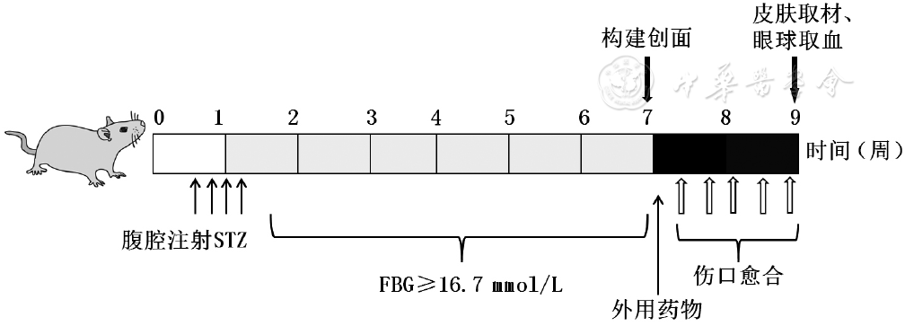
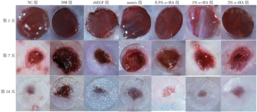
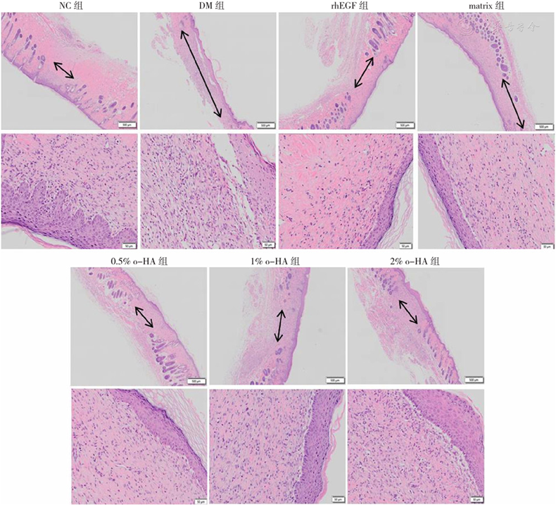
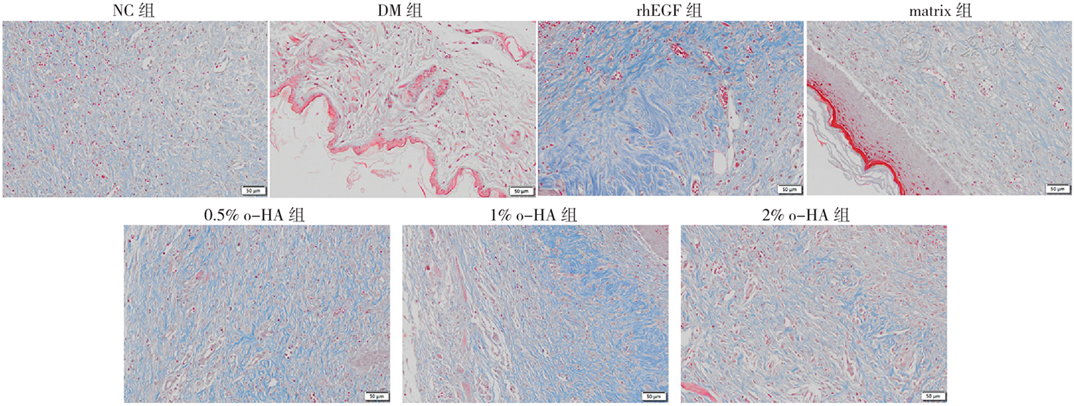
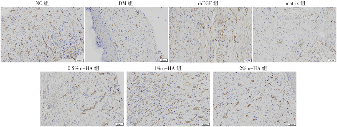
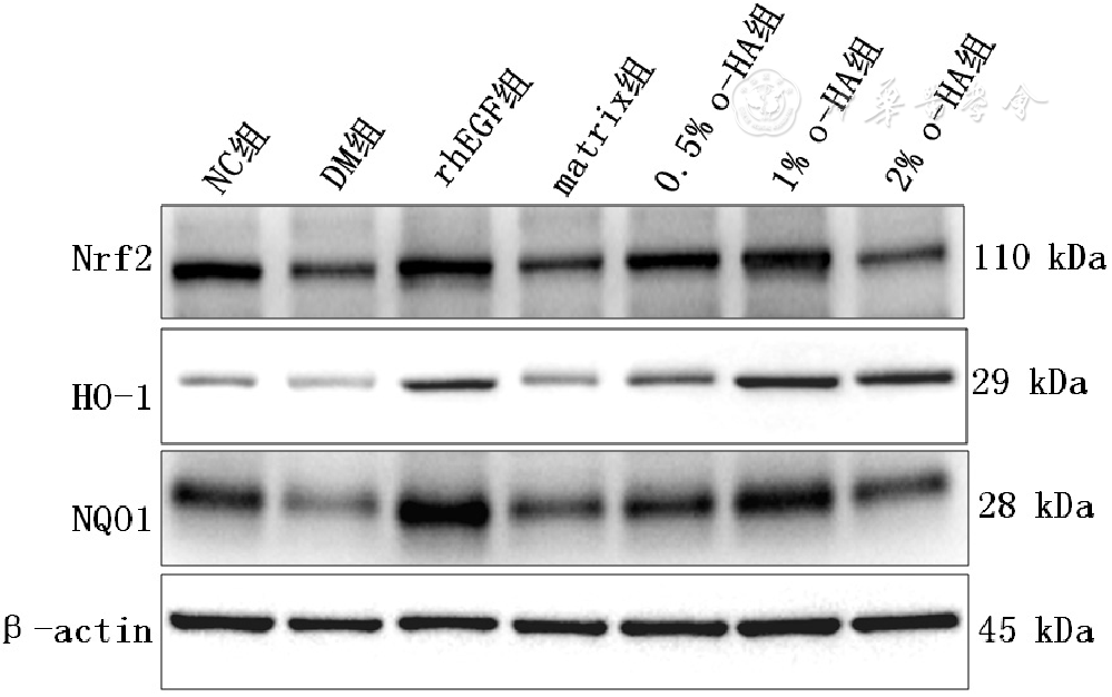
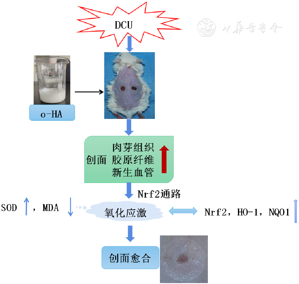
摘要: 背景 氧化应激水平过高、长期的炎症状态严重影响糖尿病患者的创面愈合,目前治疗糖尿病皮肤溃疡(DCU)的药物有生物制剂、中药、干细胞疗法等,但由于其缺乏安全性、有效性的充足证据,在临床应用中受到限制,亟需发现新的药物。透明质酸寡糖(o-HA)具有抗氧化性和抗炎性,是治疗DCU的重要方法。 目的 观察o-HA对DCU创面愈合作用并探究其对氧化应激核因子E2相关因子2(Nrf2)信号通路的影响。 方法 实验时间为2021年4—12月,取6周龄SPF级雄性昆明小鼠90只,留取10只作为正常对照组(NC组),其余经腹腔注射链脲佐菌素(STZ)诱导糖尿病小鼠模型,建模成功的60只糖尿病小鼠按照随机数字表法分为6组,每组10只,即糖尿病模型组(DM组)、重组人表皮生长因子阳性对照组(rhEGF组)、空白基质阴性对照组(matrix组)、低剂量o-HA治疗组(0.5% o-HA组)、中剂量o-HA治疗组(1% o-HA组)、高剂量o-HA治疗组(2% o-HA组)。所有小鼠利用全层皮肤切除环形夹板法建立创面模型,NC组和DM组小鼠创面不做任何处理,其他各组每日创面及创周常规消毒后,均匀涂抹各组相应药物于创面及创周,rhEGF组给药1次/d,0.5%、1%、2% o-HA组给药2次/d,连续给药14 d。治疗后第1、7、14天拍照记录DCU小鼠创面愈合情况,利用HE染色、Masson染色、CD34免疫组化染色观察给药14 d后小鼠创面肉芽组织形态变化、胶原纤维生成和新生血管,试剂盒检测给药14 d后小鼠血清超氧化物歧化酶(SOD)、丙二醛(MDA)水平,Western-blotting检测给药14 d后小鼠创面组织Nrf2、血红素加氧酶1(HO-1)、NAD(P)H醌氧化还原酶1(NQO1)的蛋白表达水平。 结果 给药第7天,rhEGF组、1% o-HA组创面愈合率高于DM组,0.5% o-HA组、2% o-HA组创面愈合率低于NC组、高于DM组;给药第14天,rhEGF组、0.5% o-HA组、1% o-HA组、2% o-HA组创面愈合率高于DM组(P<0.05)。组织形态学显示o-HA软膏干预后创面肉芽组织、胶原纤维、新生血管密度明显增多。rhEGF组、0.5% o-HA组、1% o-HA组、2% o-HA组MDA水平低于DM组,SOD水平高于DM组;0.5% o-HA组、2% o-HA组MDA水平高于NC组(P<0.05)。rhEGF组、1% o-HA组Nrf2、HO-1、NQO1蛋白表达水平均高于NC组和DM组;0.5% o-HA组Nrf2、NQO1蛋白表达水平高于DM组,HO-1蛋白表达水平高于NC组和DM组;2% o-HA组HO-1蛋白表达水平高于NC组和DM组,NQO1蛋白表达水平高于DM组(P<0.05)。 结论 o-HA对DCU小鼠创面有潜在促愈合作用,以1% o-HA软膏愈合效果最好,可加速创面闭合、再上皮化,促进血管新生,影响Nrf2途径,加速创面愈合,具有安全性高、稳定性好的优势,在临床DCU创面修复方面极具潜力。

关键词: 糖尿病, 糖尿病并发症, 糖尿病皮肤溃疡, 透明质酸寡糖, 创面愈合, 核因子E2相关因子2

Abstract:

Background

Excessive levels of oxidative stress and long-term inflammatory state seriously affect wound healing in diabetic patients. At present, there are biological agents, traditional Chinese medicines, stem cell therapy etc. used in treating diabetic cutaneous ulcer (DCU) . Due to lack of sufficient evidence of safety and effectiveness, these methods are not widely applied in clinical treatment. Thus new drugs need to be discovered urgently. Hyaluronic acid oligosaccharide (o-HA) is a potential drug for the treatment of DCU because of its antioxidant and anti-inflammatory properties.

Objective

To observe the effect of o-HA on DCU wound healing and explore its influence on oxidative stress Nrf2 signaling pathway.

Methods

This experiment was conducted from April to December, 2021. Ninety SPF male mice of six-week-old from Kunming were selected. Ten of them were reserved as the normal control group (NC group) and the rest were injected with streptozotocin (STZ) by abdomen to induce diabetes mouse model. Sixty diabetic mice modeled successfully were divided into 6 groups according to random number table with ten in each group, namely the diabetic model group (DM group) , the recombinant human epidermal growth factor positive control group (rhEGF group) , the blank matrix negative group (matrix group) , low-dose o-HA treatment group (0.5% o-HA group) , medium-dose o-HA treatment group (1% o-HA group) and high-dose o-HA treatment group (2% o-HA group) . The DCU model was established in all mice by way of the full-thickness skin excision ring splint. The wounds in NC and DM group were not treated. For the other groups, the corresponding medicines were evenly applied to and around the wounds after daily disinfection. The mice in rhEGF group were administered once a day while the mice in the 0.5%, 1% and 2% o-HA group were medicated twice a day for consecutive 14 days. The wound healing condition of DCU mice was recorded after being treated on the first, the 7th and the 14th day. The morphological changes of granulation tissue, collagen fiber formation and angiogenesis in mice wounds were observed by HE staining, Masson staining and CD34 immunohistochemical staining. The serum SOD and MDA levels of mice were examined by the kits. The Nrf2, HO-1 and NQO1 protein expression in wound tissue of mice were detected by Western-blotting after the administration of 14 days.

Results

On the seventh day of administration, the wound healing rate of rhEGF group and 1% o-HA group was higher than that of DM group and the wound healing rate of 0.5% o-HA group and 2% o-HA group was lower than that of NC group and higher than that of DM group. On the 14th day of administration, the wound healing rate of rhEGF group, 0.5% o-HA group, 1% o-HA group and 2% o-HA group was higher than that of DM group (P<0.05) . The histomorphology showed that the granulation tissue of the wound, collagen fiber and density of neovascularization were significantly increased after o-HA ointment intervention. The level of MDA in the rhEGF group, 0.5% o-HA group, 1% o-HA group and 2% o-HA group was lower than that of DM group, and the level of SOD in these four groups was higher than that of DM group; the level of MDA in 0.5% o-HA and 2% o-HA groups was higher than that of NC group (P<0.05) . The levels of Nrf2, HO-1 and NQO1 protein expression in both rhEGF group and 1% o-HA group were higher than those of NC and DM group; the levels of Nrf2 and NQO1 protein expression in 0.5% o-HA group were higher than those of DM group, and the level of HO-1 protein expression was higher than that of NC and DM group; the level of HO-1 protein expression in 2% o-HA group was higher than that of NC and DM group, and the level of NQO1 protein expression was higher than that of DM group (P<0.05) .

Conclusion

o-HA has a potential healing effect for DCU wound in mice, and the healing effect of 1% o-HA ointment is the most remarkable. It can help wound healing by accelerating wound closure and re-epithelization, promoting angiogenesis and affecting the Nrf2 pathway. With advantages of high safety and good stability, o-HA has great potential in clinical diabetic wound healing.

Key words: Diabetes mellitus, Diabetes complications, Diabetic cutaneous ulcers, Hyaluronic acid oligosaccharides, Wound healing, NF-E2-related factor 2